Like the vast majority of white birches in this area, this one of mine was dying two years
ago - its leaves were turning yellow and dropping off.
However, the tree miraculously recovered after a fungicide spraying.
While the first half of all the branches died, the outer half was saved and now appears very healthy.
The following pictures were taken in late May, about a week before a major spore
release.
1
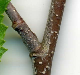
The branch junction shown here is about a foot from the end of a healthy branch.
Its bark appears healthy and very few spores are present at the junction.
2
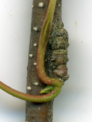
This leaf stalk is about a foot closer to the trunk, and still on new growth.
There are very few spores present.
3
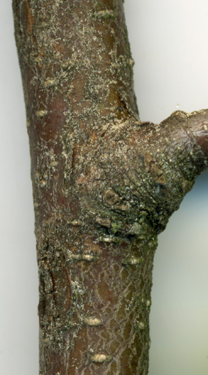
In contrast, here we see a section of the branch about 4' from the branch tip.
There were a few dead twigs coming off this branch a few inches further toward this branch's tip.
After that, all the twigs were alive.
So this wood was probably created just before the fungicide spraying about two years
ago - hence the high spore density at the branch junction.
With all those spores present, one would think that this tree should have a
severe infection of white canker. Of course, to confirm a white canker
infection, one needs more than tan dots. The following pictures were taken in
early October using a 400x digital microscope. At this time the leaves were
beginning to turn color and fall from the trees. The first two pictures below were from
the bark of a twig about 3/16 inch in diameter. The third picture was of a leaf
bottom.
4
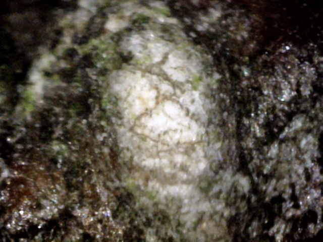
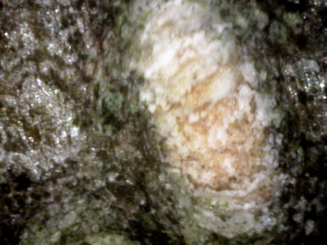
The bark shows few typical white canker objects.
However, there are these numerous erupting white oval objects covered in what looks
like crystalized snow.
5
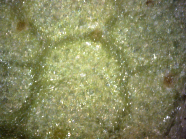
The bark is scattered with these oval erruptions.
Each consists of particles of white canker on top, with tan wound wood showing
through underneath.
6
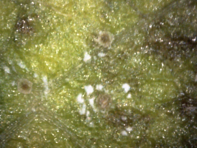
Generally a very boring leaf bottom!
The only thing of note is the light sprinkling of these amber circles.
Sometimes the top surface of a leaf can display white canker disease symptoms.
The following three photos show the the leaf top, although there's not much to see!
7
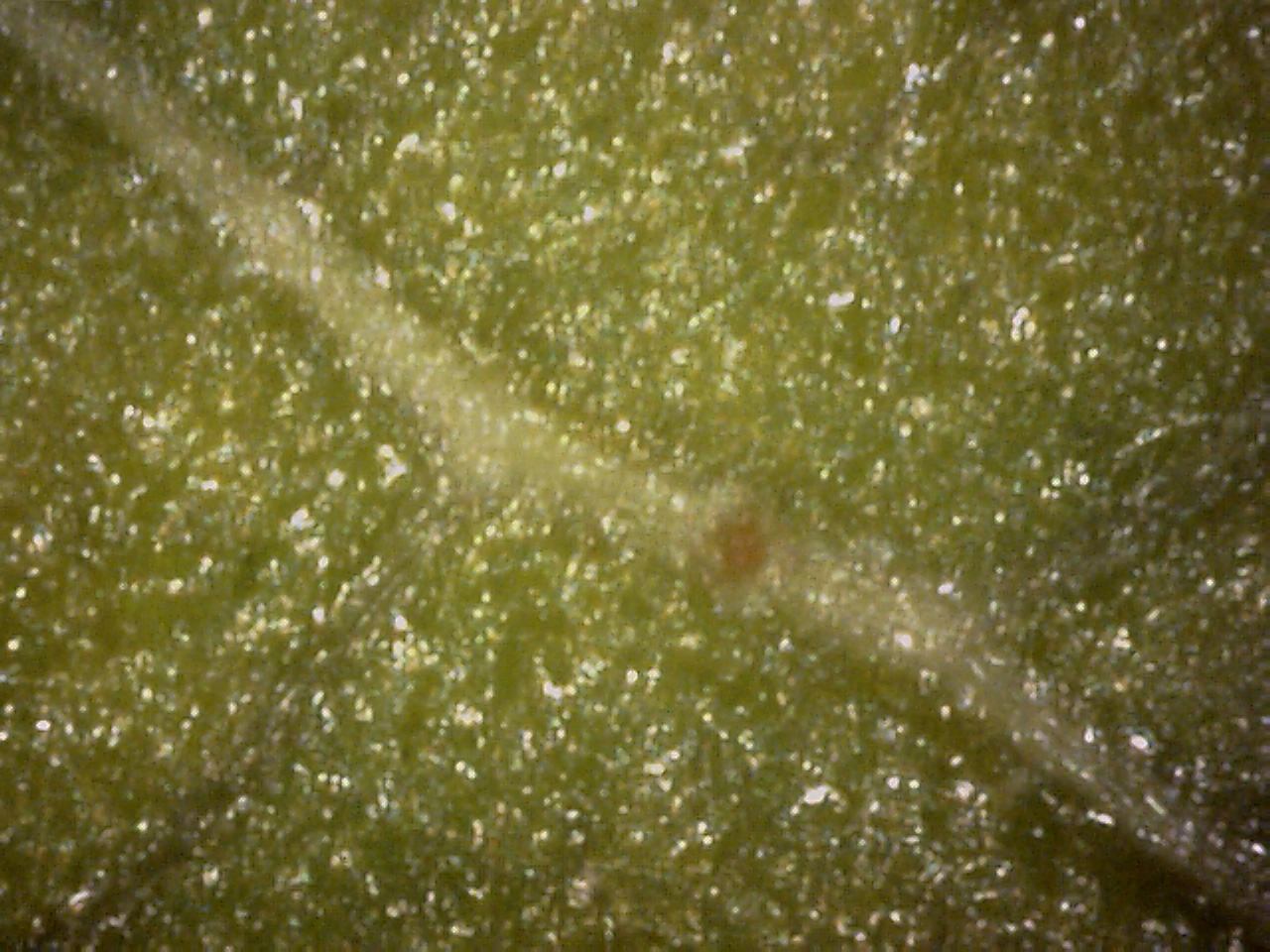
The leaf top is sprinkled with these brown to amber spots.
These spots seem to prefer leaf veins, and usually have canker material growing
around them, as shown here.
8
As usual, this amber spot sits directly on a leaf vein.
But while veins are normally green, this particular vein seems to have been
taken over by white canker.
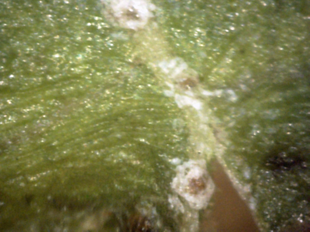
9
The association between the amber spots on the leaf top and white canker
is particularly strong here. The combination resembles a fried egg - sunnyside up.
If you can't see anything of interest on the surfaces of the leaf, a look inside the leaf
often proves more helpful. The following three pictures show cross-sections of the leaf, made
by slicing a leaf with a razor and then examining the edge with a digital microscope at 400x.
 ←
←
10
It now becomes more evident that the amber leaf spots seen so frequently on the top
and bottom surface of the leaf are strongly associated with white canker.
Here the amber spot has canker on the left, right, and especially the bottom/leaf interior.
There is also a transparent tube of canker on the left, running from the top to
the bottom of the leaf.
 ↓
↓
11
Here is another one of those amber spots on the leaf top (red arrow).
It is raised above the surface, and seems to have an extensive canker root system.
The razor making this cross-section went from right to left.
Therefore, the gap to the right of the canker once again seems to indicate that the
canker material is relatively firm - at least more cohesive than the surrounding leaf tissue.
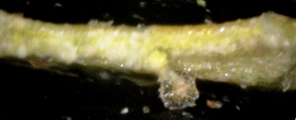 ←
↑
←
↑
12
This (red arrow) appears to be an amber spot on the bottom surface that was was torn downward
by the razor making the leaf cross-section.
It looks like the amber spot was closely allied with the canker which enveloped
the leaf cells on the bottom surface and killed them, turning them transparent (you could
say they were turned into a ghost!).
It also looks like the amber spot is surrounded by bubble-wrap!
Note that the leaf bottom is free of this blubble-like material where it was torn
off but adjacent areas still have it.
Also, check out the amber spot on the left (blue arrow) - it is more like a raised dome rather
than the surface of sphere.
It's puzzling as to why white canker would go through the trouble to create these amber
spots on the leaves when the leaves are only days away from dying and dropping off the tree.
An alternative explanation is that the leaf creates these spots in response to a strong
infection, in an attempt to wall off the infection, much as wound wood is used to close up
a breach of the bark. Maybe that's why these spots are the color of wound wood.
The final set of pictures shows cross-sections of a 3/16 inch twig. Even though this is a
very small part of the tree, if white canker is present, it shows up well in twigs. A
nice side benefit is that twigs of this size are very easy to obtain using just a
pruning clippers. Once again, all pictures were taken with a digital microscope at 400x.
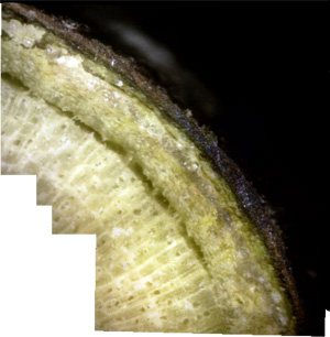
13
This is a photomerge of 5 pictures to show internal fracturing.
The canker growing in the phloem is causing it to expand, causing an air gap to
form between the xylem and phloem, which is being bridged by a few rays. The
side benefit for the tree is that the canker is having a more difficult time
jumping to and infecting the xylem.
14
This is the most "normal" view of the entire twig cross-section.
Still, blobs of white canker can be seen growing in the phloem, and the bark is beginning
to separate at the far left and far right.
In addition, white canker can be seen growing on the outside of the bark.
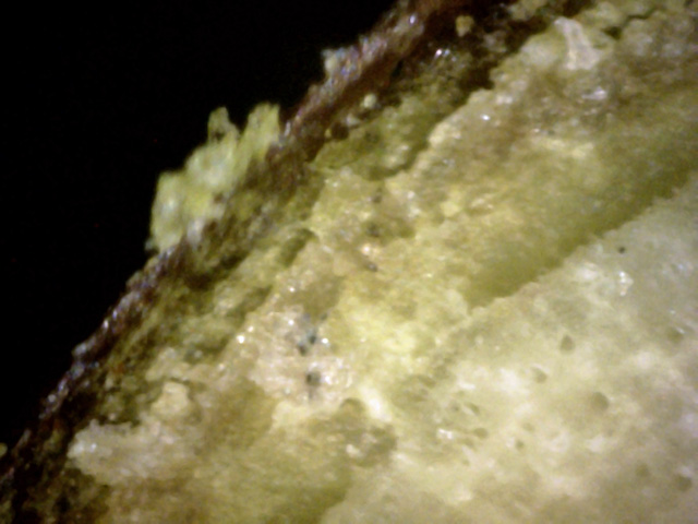
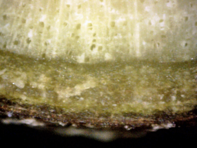
15
There is extensive white canker present in the phloem in this view.
White canker sits on the bark surface, and the destruction of the dark green cortex
has left a void under the bark.
 →
←
←
←
←
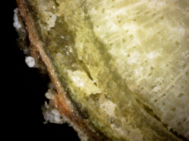
16
A good example of the destruction of the outer phloem (just under the dark-green cortex
layer, which in turn sits under the bark) by white canker.
But the especially interesting feature is a spore of white canker sitting on the outside
of the bark (red arrow).
Note that directly under this spore the outer bark (blue arrow) has been completely replaced
with canker material.
And, the white spore substance above and below it are both sitting above their own resevoir
(green arrows) of gray canker material under the bark.
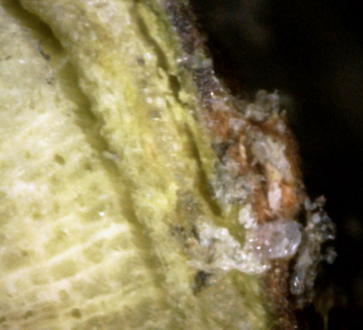
17
A good example of a battle between the bark of a tree and the white canker
trying to destroy it. The bark is trying to grow wound wood around the canker in
an attempt to heal the infection. But it looks like the canker is fighting back,
creating extensive canker material on both sides of the wound wood.
In summary, at this time of the year it was very difficult to diagnose white canker with the
unaided eye. Using a 400x microscope, the bark was only a little helpful in diagnosis.
Views of the upper and lower part of infected leaves likewise weren't too helpful.
However, leaf cross-sections showed that a good amount of white canker was present.
But the best evidence of all once again came from a twig cross-section, where there was
clear evidence of widespread white canker growth under the bark, specifically at the
junction between the cortex and phloem.